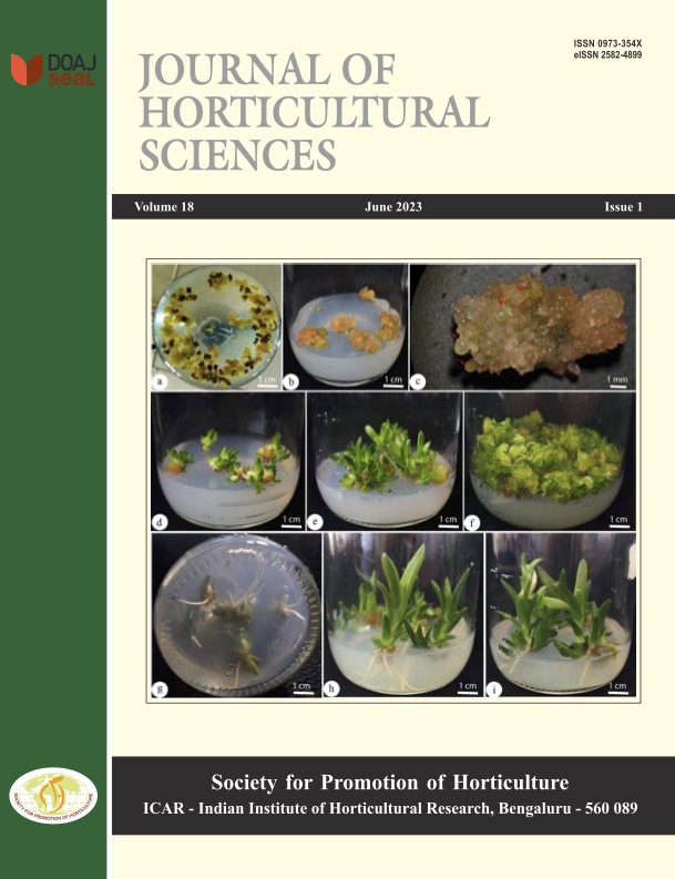
Standardization of sterilization protocol for explants and its suitability for direct organogenesis in tuberose cv. Arka Vaibhav
DOI:
https://doi.org/10.24154/jhs.v18i1.2160Keywords:
Aseptic culture, direct organogenesis, explants, surface sterilizationAbstract
A study was carried out to standardize the sterilization protocol for different explants (terminal stem scale,
immature flower bud and tepal segment) and to select the suitable explant for the direct organogenesis of tuberose cv. Arka Vaibhav. The highest survival per cent (100) and uncontaminated cultures (0.00) of terminal stem scale explant was observed in pre-treatment with overnight soaking of terminal stem scale in the solution comprising carbendazim (0.1%), chlorothalonil (0.05%) and myristyl trimethyl ammonium bromide (cetrimide) (0.05%) and subsequently surface sterilization with 70% ethanol (1 min), 4% sodium hypochlorite (10 min) followed by 0.1% HgCl2 (15 min). The explant immature flower bud recorded the highest survival per cent (100) and maximum aseptic cultures in the treatment T1 comprised of 1.0 drop Tween-20 + 70% ethanol (30 sec) and 1% sodium hypochlorite (3 min). Pre-treatment of tepal segment explant in 0.1% carbendazim (30 min) solution followed by surface sterilization with combination of 1.0 drop Tween-20 + 70% ethanol (30 sec) followed by 1% sodium hypochlorite (3 min) registered 91.66% of survival with the minimum contamination (10%) in the treatment. Among the three explants used, the terminal stem scale was found suitable for direct organogenesis with early greenness (5.72 days) and highly responsive to shoot induction (100%) in MS medium supplemented with 4 mg/L BAP + 0.1mg/L IAA. Other two explants viz., immature flower bud and tepal segment failed to respond for direct organogenesis by shoot induction instead produced profuse callus.
Downloads
References
Anonymous, 2023. Indiastat; 2021-22 3rd advance estimates. https://www.indiastat.com/table/ agriculture/area production-tuberose-india- 2011-2012-2021-2022/964510.
Aslam F., Naz S., Tariq A., Ilyas S. and Shahzadi K. 2013. Rapid multiplication of ornamental bulbous plants of Lilium orientalis and Lilium longiflorum. Pak. J. Bot., 45(6): 2051-2055.
Bora, S. and Paswan, L. 2003. In vitro propagation of Heliconia psittacorum through axillary bud. J. Ornam. Hortic., 27(5): 450-452.
Chen, L., Zhu X., Gu, L. and Wu, J. 2005. Efficient callus induction and plant regeneration from anther of Chinese narcissus (Narcissus tazetta L. var. Chinensis Roem). Plant Cell Rep., 24(7): 401-407.
Copetta, A., Marchioni, I., Mascarello, C., Pistelli, L., Cambournac, L., Dimita, R. and Ruffoni, B. 2020. Polianthes tuberosa as Edible Flower: In vitro propagation and nutritional properties. Int. J. Food Eng., 6(2): 57-62.
Dilta, B. S., Sehgal, O. P., Pathania, N. S. and Chander, S. 2000. In vitro effect of NAA and BA on culture establishment and bulblet formation in lily. J. Ornam. Hortic., 3(2): 65-70.
Gajbhiye, S. S., Tripathi, M. K., Vidya Shankar, M., Singh, M., Baghel, B. S. and Tiwari, S. 2011. Direct shoot organogenesis from cultured stem disc explants of tuberose (Polianthes tuberosa Linn.). J. Agric. Sci. Technol., 7(3): 695-709.
Jyothi, R., Singh, A. K. and Singh, K. P. 2008. In vitro propagation studies in tuberose (Polianthes tuberosa Linn.). J. Ornam. Hortic., 11(3): 196-201.
Kadam, G. B., Singh, K. P., Singh, A. K. and Jyothi, R. 2010. In vitro regeneration of tuberose through petals and immature flower buds. Indian. J. Hortic., 67(1): 76-80.
Kanchana, K., Kannan, M., Hemaprabha, K. and Ganga, M. 2019. Standardization of protocol for sterilization and in vitro regeneration in tuberose (Polianthes tuberosa L.). Int. J. Chem. Stud., 7(1): 236-241.
Krishnmurthy, K. B., Mythili, J. B. and Meenakshi, S. 2001. Micropropagation studies in single vs double types of tuberose (Polianthes tuberosa L.). J. Appl. Hortic., 3(2): 82-84.
Leifert, C. and Cassells, A. C. 2001. Microbial hazards in plant tissue and cell cultures. In vitro Cell. Dev. Biol. Plant., 37(2):133-138.
Mir J. I., Ahmed N., Itoo H., Sheikh M. A., Rizwan R. and Wani S. A. 2012. In vitro propagation of Lilium (Lilium longiflorum). Indian J. Agric. Sci., 82(5): 455-458.
Mishra, A., Pandey, R. K. and Gupta, R. K. 2005. Micro-propagation of tuberose (Polianthes tuberosa L.) cv. Calcutta Double. Prog. Hortic., 37(2): 331-335.
Pandey R. K., Singh A. K., Mamta S. 2009. In vitro propagation of Lilium. Biol. Forum-An Int. J., 1(2): 26-28.
Rather, Z.A., 2010. Studies on in vitro propagation of Peony (Paeonia spp.). Ph.D. Thesis submitted to Sher-e-Kashmir University of Agricultural Sciences and Technology, Kashmir, Shalimar.
Ray, S. S. and Ali, N. 2016. Biotic contamination and possible ways of sterilization: A review with reference to bamboo micropropagation. Braz. Arch. Biol. Technol., 59: e160485.
Sangavai, C. and Chellapandi, P. 2008. In vitro propagation of a tuberose plant (Polianthes tuberosa L.). Electronic J. Biol., 4(3): 98-101.
Surendranath, R., Ganga, M. and Jawaharlal, M. 2016. In vitro propagation of tuberose. Environ. Ecol., 34(4D): 2556-2560.
Taha L. S., Sayed S. S., Farahat M. M., El-Sayed I. M. 2018. In vitro culture and bulblet induction of Asiatic hybrid lily ‘Red Alert’ J. Biol. Sci., 18(2): 84-91.
Downloads
Published
Issue
Section
License
Copyright (c) 2023 Mahananda Patil , T U Bharathi , T R Usharani , Rajiv Kumar , B S Kulkarni

This work is licensed under a Creative Commons Attribution-NonCommercial-ShareAlike 4.0 International License.
Authors retain copyright. Articles published are made available as open access articles, distributed under the terms of the Creative Commons Attribution-NonCommercial-ShareAlike 4.0 International License, which permits unrestricted non-commercial use, distribution, and reproduction in any medium, provided the original author and source are credited. 
This journal permits and encourages authors to share their submitted versions (preprints), accepted versions (postprints) and/or published versions (publisher versions) freely under the CC BY-NC-SA 4.0 license while providing bibliographic details that credit, if applicable.


 .
. 


